Monday, February 28, 2011

Wuchereria bancrofti: An Elephant of a Problem

[5]
Introduction:

Wuchereria bancrofti is a thread-like nematode that is one of the three different nematodes that cause Lymphatic filariasis in humans. Today, there are 120 million people infected with these parasites. These parasites can be found in 80 countries: in the humid and tropical areas (where mosquitos are apparent) of Africa, Asia, isolate America areas, and the Pacific islands. Usually in areas of poverty. The symptoms of this parasite are usually asymptomatic. This parasite will not cause harmful symptoms until the body reacts and the lymphatic tissues (usually in the groin and the legs) begin to swell. Often, the infected that are left untreated will develop elephantiasis in the legs and groin region. This parasite itself is not deadly, but left untreated can cause deadly infections and can ultimately hinder a person mobility[1].


Symbiont Description:

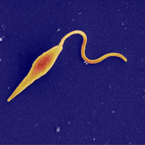
The Wuchereria bancrofti are thread-like nematodes. The female nematodes are normally 10 cm long and .2mm wide while the males are 4 cm long. These organisms thrive in the hosts lymphatic system and can produce around 50,000 microfilaria a day [2]. These microfilaria leave the lymphatic system and circulate the body. During the day, they thrive in the lungs and at night, they sense their hosts body temperature drop, and travel and emerge in the peripheral veins/arteries at night in order to spread through a vector, typically a mosquito [3]. Wuchereria bancrofti can live as in the body as microfilaria for up to 12 months. It takes about 6-12 months for Adult worms to develop from the larval stage and can live up to 4-6 years [2].

Female W. bancrofti [6] Male W. bancrofti [6]

Host Description:

These nematodes usually begin transmission through a mosquito vector and transmission is completed when the mosquito bites a human. Typically, mosquitos bite during the night in humid areas. Humans are the only known host. [3]


Life Cycle:

[4]
As previously stated, the Wuchereria bancrofti has two hosts: a mosquito and a human. The CDC has listed the mosquito as being the first step of this nematodes lifecycle. The first part of their lifecycle occurs when an infected mosquito bites an innocent standby for a lovely meal. The third stage larvae will enter into the human through the opened bite made by the mosquito. It is then that these larvae make their way to the lymphatic tissues where they will develop into adults [4]. This can take up to 12 months [1]. The adults will then mate and create up to 50,000 microfilariae a day [1]. As a 'new born' the microfilariae are sheathed. As stated previously, during the day, the microfilariae are found in the lungs and during the night, they sense a change in their hosts' body temperature and will migrate to the peripheral veins/arteries where they await to be found by a hungry mosquito [4]. They follow the hosts sleep cycle as they migrate to the peripheral veins/arteries [8]. The mosquito will unknowingly consume the microfilariae during their meal. Once inside the mosquito, the microfilariae will then loose their sheaths where they will find their way to the mosquitos midgut and their thoracic muscles. In 1-2 weeks, they will turn into first stage larvae and then gradually develop into a third-stage infection larvae where they are then able to infect another host, thus the life cycle restarts [4].

Ecology:

This parasite only has two hosts: human and mosquito (vector). It is prevalent in humid climates and where mosquitos actively are. As mentioned before, these areas are the humid and tropical areas (where mosquitos are apparent) of Africa, Asia, isolated areas in the Americas, and the Pacific islands [1]. The bottom map shows these particular areas that are affected with this parasite.

[6]

Today, 120 million people today are affected by this parasite. Even more devastating, more than 30% of this infected populace are severely incapacitated by the horrendous disease this parasite causes. In the 80 countries this parasite is associated with, 1 billion people are at risk for being infected [1]. This disease is diagnosed with a simple test. An antigen-detection test, known better as the ITC test can scan blood sample and will reveal the results within minutes. The treatment of these parasites and the disease that it causes is three anti-parasitic drugs, albendazole and mectizan, and the possible surgical removal of the swollen limbs. Even then, the people affected with this disease aren't able to get back their mobility. The Global Alliance to Eliminate Lymphatic Filariasis is an active partner to the World Health Organization (WHO) whom are actively trying to rid the world of this disease. Several feats that they are implementing is having mass drug administration of this parasites killer Albendazole, Mectizan, and DEC. The latter drug has one set back: It cannot be used in African countries. When using this drug, severe side reactions can occur when other infections of the body, such as river blindness, are present. Instead the first two drugs are more commonly used [7]. Both American drug companies, Glaxo Smith Kline and Merck, are committed to fund free medication for this eradication program [8]. Other ways this group is fighting this parasite is through passing out mosquito bug nets, to prevent mosquito attacks [7]. Below is a video of this movement.


[8]

The Wuchereria bancrofti is a perfect example of circadian periodicity and emerging when their 2nd host, the mosquito, feeds [5]. The microfilariae senses a change in their hosts body temperature and will make way to their hosts peripheral blood vessels and remain there until they sense another change in body temperature and will then return back to the lungs [1]. As mentioned before, the microfilariae move in rhythm with their hosts sleep cycle [8]. While in the peripheral blood vessels, the microfilariae hope to get picked up by the 2nd host, the mosquito, where they will then grow and develop into their infectious larval stage and will hopefully reach another human host [1].

Below is a video describing this parasites life cycle.

[9]

Sources:
1. http://www.parasitesinhumans.org/wuchereria-bancrofti-lymphatic-filariasis.html
2.http://www.phac-aspc.gc.ca/lab-bio/res/psds-ftss/wuchereria-bancrofti-eng.php
3. http://animal.discovery.com/invertebrates/monsters-inside-me/lymphatic-filariasis-w-bancrofti/
4. http://www.dpd.cdc.gov/dpdx/html/frames/a-f/filariasis/body_Filariasis_w_bancrofti.htm
5. http://emedicine.medscape.com/article/996732-overview
6.http://www.science.smith.edu/departments/Biology/SWILLIAM/fgn/pnb/wuchban.html#path>pathologies described below.
7. http://www.filariasis.org
8. http://www.youtube.com/watch?v=dnWwHthkGkY&feature=player_embedded
9.http://animal.discovery.com/videos/monsters-inside-me-the-filariasis-parasite.html
Naegleria fowleri: Road to Losing Your Mind, Literally!

Introduction

Have you ever gone swimming in any warm freshwater lake, river, or hot springs recently? Perhaps Lake Lanier? If your answer is yes then I am sorry to inform you but there is a chance you are infected with Naegleria fowleri (N. fowleri)! Better act quickly because this infection is deadly and usually causes death within 1-12 days! However, before I scare you too much, there is some good news. Our body does a pretty good job of fighting the encounter with the parasite before it can actually lead to infection. Between the years 2000 to 2009 only 30 infections were reported in the U.S [1]. That is a low number considering the high exposure one has with this interesting parasite.
It is very common but rarely causes disease. N. fowleri is a free-living ameba that is found in warm freshwaters all around the world. The primary disease caused when infected is primary amebic meningoencephalitis (PAM). It is very rare but an extremely serious disease with a 95% mortality rate. There are several treatments that include: amphotericin B, rifampicin, and miconazole [2]. Unfortunately almost all patients die. Image: [3]

Symbiont Description

N. fowleri is a member of the subphylum Sarcodina and superclass Rhizopodea. The superclass Rhizopodea includes several species of amebas. The genus Naegleria includes several species, however, only N. fowleri is known to produce human disease [2]. N. fowleri is a free-living organism, meaning that they can survive without a host. This is a great advantage to the parasite and explains why their attacks are fatal [4]. Hosts are necessary for survival of parasites that are not free-living. They have to think twice before killing the host. N. fowleri, on the other hand, can survive without a host and so do not have to avoid killing them.

An interesting aspect of this parasite is that it has both a flagellated and unflagellated form. More specifically it has two flagella. During the flagellated stage it does not feed. In laboratory tests, N. fowleri loses the flagella when placed in distilled water. The ameba gains the flagella again when there is a change in the environment. During unfavorable conditions the amoeba can also form cysts and this allows it to survive harsh conditions. Image: [5]
Host Description

For parasites like N. fowleri that thrive in oxygenated environment, the brain, which is oxygenated tissue, is the perfect home. However, finding a way into this nurturing environment is a challenge.

The brain has an especially more protective barrier called the blood-brain barrier.This barrier is made of cells that make a tight seal along blood vessels that prevent mostly everything from the bloodstream, which includes brain parasites like N. fowleri, from leaking through [6]. Unfortunately for us, N. fowleri rise up to the challenge very well. So how do they enter the brain? [Image [7]

The attack:

The parasite attaches to the inside of the host’s nose and follows the olfactory nerve path to reach the brain. When it has reached the brain, the damaging effects commence. N. fowleri crosses the barrier by actually eating the nerve cells.This is obviously very bad for the host and is the main reason for the rapid death. The parasite has surface proteins that allow it to cut a hole in the covering of the cell. When it pierces the hole the contents of the neuron leak out and the parasite feed on the nutrients it contains. Furthermore, this special parasite has proteins that are able to sense the presence of certain nutrients, and this causes it to send signals to the rest of the cell indicating in which direction the parasite should move to eat those nutrients [6].

The human body cannot detect the intrusion of the parasite in the brain because the parasite is able to avoid detection by another set of proteins called CD59. This prevents the parasite from being taken up by phagocytes or being destroyed [4]. In addition, there are other proteins on the surface that direct the parasite to the most vulnerable areas of a neuron [6].Basically this parasite has evolved its own GPS system of the human brain! What can our body do when our mind is being eaten alive? Image: [8]

Well the human body does a superb job of preventing the parasite from even getting so close to the brain and it does so by using snot! A primary defense of the body is to secret mucus so that the parasite cannot adhere and move up the nose. N. fowleri again rises up to this challenge by producing enzymes to digest the mucus, this process is called mucinolytic activity [6]. In the competition of mucus production versus the mucinolytic enzyme effectiveness the body does an exceptional job of trapping the parasites. This is supported based on how often we are exposed to this parasites and how often they actually cause infection.

Life Cycle

The life cycle of N. fowleri has 3 stages. In the ameboid trophozite stage, the parasite feeds on bacteria and replicates through binary fission where the nuclear membrane remains intact. Replication can only occur in this stage. This is also the stage where human infection can occur. When there is a change in ionic concentration the

trophozite changes to a flagellate form. This is reversible and they will change back when conditions are back to normal. During the flagellated form, the parasite does not infect humans. When water temperature decreases and become very unfavorable N. fowleri encyst and this allows them to survive the winter until the next summer comes around [9]. Image:[10]

Ecology

N. fowleri is a free-living amoeba found all around the world. They live anywhere there is warm freshwater, like lakes, rivers, or geothermal (naturally hot) water sources. N. fowleri can also be found in swimming pools that are poorly maintained. This parasite is not found in the ocean or any other salt waters. Most of the infections have occurred during the summer months of July, August, and September and mainly in the southern states [1]. One of the reasons it has not become more prevalent is that it cannot be spread from person to person [11]. It is only spread through nasal contact with water infected with the amoeba. N. fowleri causes PAM, which is a brain infection that leads to the destruction of brain tissue. Their symptoms are similar to bacterial meningitis in its early stages. Aggressive treatments with amphotericin B and miconazole have proven positive for effectiveness in labs, however again the treatment and diagnosis is 95% of the time too late. With proper education to the public it can be caught early enough to use the treatment options [2].


Some signs and symptoms of infection include stiffness, cramping and pain in the muscles of the back and neck. Weakness, severe headaches, seizures, nosebleeds, rapid and shallow breathing, swollen and painful lymph nodes is possible. Decreased appetite, loss of smell and problems tasting food, confusion, hallucinations, and difficulty with concentration may also be associated. [11]. Video: [12]

An Example of the Host’s Two Lines of Defense

N. fowleri’s host, the human body, displays the perfect example of the concept of a host having two lines of defense.The first line of defense is avoiding the parasite by appropriate behavior. If the encounter still does take place, the second line of defense is immunity, which is basically to expel the intruder.

Humans have the advantage of avoiding the parasite just by having the ability to read and make signs. Freshwater bodies of water more susceptible to N. fowleri have signs and display warnings to recreational water users to beware of the ameba. Keeping the nose plugged with nose plugs is just one simple tactic to avoid an encounter with this brain-eating parasite. If the encounter does take place, the body has internal defenses (the second line of defense) such as increasing mucus production that prevents the parasite from moving up the nose and reaching the brain. However, the host is good as dead if the parasite is able to pass this line of defense. Once it has reached the brain, N. fowleri begins the prompt degeneration of brain tissue and rapid death follows [6].

So the next time you decide to go swimming in any warm freshwater lake make sure you bring nose plugs. You do not want Naegleria fowleri swimming up your nose and eating your brain! Image: [13]

References

[1]http://www.cdc.gov/parasites/naegleria/faqs.html

[2]http://emedicine.medscape.com/article/972044-overview

[3]http://www.flickr.com/photos/trenchcoatjedi/2285088742/

[4]http://www.bio.davidson.edu/people/sosarafova/Assets/Bio307/alkim/Evasion%20of%20the%20immune%20system.html

[5]http://en.academic.ru/dic.nsf/enwiki/288084#

[6]http://eands.caltech.edu/articles/LXVI4/brainworms.html

[7]http://www.colorado.edu/intphys/Class/IPHY3730/05cns.html#outline

[8]http://infomationhealth.blogspot.com/2010/10/living-creatures-that-can-live-in-our.html

[9]http://www.stanford.edu/class/humbio103/ParaSites2004/N.%20Fowleri/life%20cycle%20&%20morphology.htm

[10]http://www.ehagroup.com/resources/pathogens/acanthamoeba-spp-and-naegleria-fowlerimeningitis/

[11]www.polk.wateratlas.usf.edu/upload/documents/fact_sheetAmoebae.pdf

[12]http://health.discovery.com/videos/monsters-inside-me-brain-eating-amoeba.html

[13]http://casperswanders.blogspot.com/2010/11/nevada-desert-adventure-nanfa_12.html


Sunday, February 27, 2011

Philophthalmus gralli: A real eye-opener


Introduction:
Philophthalmus gralli is often referred to as the oriental avian eye fluke. It was once thought that this parasite was only found in the eastern part of the world but we now know that the parasite is found on almost every continent. P. gralli is found in most bird species ranging from ducks to pigeons to ostriches. The parasite is ingested through the mouth of a bird that feeds on aquatic vegetation. From there the parasite travels to its final resting spot in the conjunctival sac of the bird’s eye. Once the parasite is in the eye, the bird will experience severe watery discharge from the infected eye. Other common signs of philophtalmiasis include swollen eyes or a mild edema. Humans may also be infected by this parasite, but they are considered an accidental host. However, human cases have been found in the United States, Central Europe, the Middle East and Southeast Asia and Japan. [1]


Symbiont Description:

The oriental avian eye fluke is part of the genus philophthalmus and of the species gralli. P. gralli takes many different shapes throughout its life. When it finally reaches its adult stage, the fluke is fusiform in shape and is covered with spines. The body is elongated and rounded towards its posterior end. The trematode can grow to a size between 2.5 – 3.3 mm long and .5 – 1.2 mm wide. The parasite contains two suckers that help it travel though the body and against the flow of blood. The reproductive organs of both the male and female are found within the fluke itself in separate compartments. [5] [6]


Host Description:
Philophthalmiasis gralli is capable of infecting millions of avian species. The parasite infects its first intermediate host: a snail. Thiara spp. and Melanoides spp. are believed to be two species of snails that are known to carry the parasite. From there the parasite reaches its finial host. The final host can be any number of birds. Research has shown an infection in ostriches in Zimbabwe [2] and baby ducks in Jordan [4] but those are not the only cases. An accidental host includes Humans, although this is not common. [1]


Life Cycle:


1.) The definitive host sheds the fully-embryonated egg into water by means of flushing the eye with water if it is a human host or by the body’s immune system causing the eye to water in avian hosts.
2.) Once in water the miracidia hatch immediately and penetrate the snail by two glandular ducts on each side of the apical gland
3.) The miracidia inside the snail turn into cercariae
4.) Snails release the cercariae and they began to form cysts on aquatic vegetation
5.) A wild bird or human becomes infected with the parasite when they ingest the metacercariae
6.) Inside the host’s mouth the cysts open and the parasite travels to the eye using its two suckers
7.) In the host’s eye the parasite lives until it is flushed out with water and then starts the cycle again [1] [5]

Ecology:
Philophthalmus gralli is mostly found in wild birds and the prevalence rate is different depending on specie type and country. The parasite can be found anywhere you find birds and so it is prevalent worldwide. The parasite has only been found in humans in a few countries including United States, Central Europe, the Middle East and Southeast Asia and Japan. The parasite is shown to cause blindness in birds if left untreated and it is not known what it does to humans. It is also unclear why the parasite choices the eye of the bird since the bird is a terminal host and will most likely not be eaten by anything other. It is also unclear as to what the parasite lives off in the eye. The parasite can be treated by flushing the eye out with water and because it is so easy to treat it is not likely to kill its host. There are currently no medications that can be taken to ride the human eye of the parasite. Economically the parasite can cause infections in ducks and chickens, which are consumed by humans. The infection can lower the number of chickens being sold and eventually if the number of infected chickens gets high enough the price of chicken may increase as well. [1] [3] [5]


An example of __________:
The parasite Philophthalmiasis gralli is a perfect example of how having more intermediate hosts’ increases the likely hood of infecting the final host. The snail being an intermediate host allows more parasites to infect it and thus the snails can provide a home for the parasites to grow in. The larger number of snails also allows for a large number of cysts to be added to the aquatic vegetation. This means that a single blade of grass can be infected with more cysts, which provided a better chance that the wild bird that eats that plant get infected.


References:
[1] http://www.dpd.cdc.gov/dpdx/html/Frames/M-R/Philophthalmiasis/body_Philophthalmiasis_page2.htm#Clinical%20Features
[2] http://www.ncbi.nlm.nih.gov/pubmed/16300188
[3] http://www.jstor.org/pss/2423036
[4]http://journals.cambridge.org/action/displayAbstract;jsessionid=4E1C7690F476EAF2BD154F0F21561E13.tomcat1?fromPage=online&aid=1719072
[5] http://www.biologia.ucr.ac.cr/rbt/attachments/volumes/vol50-2/19-D%C3%ADaz_Experim.pdf
[6] http://en.wikipedia.org/wiki/Philophthalmus_gralli

Thursday, February 24, 2011

Acanthamoeba: You Won’t Even See It Coming

Acanthamoeba: You Won’t Even See It Coming
My face frozen in a contorted grimace, I could still make out the blunt steel needle closing in on my eye. 'Keep still, and open your eye as wide as you can,' said the doctor, as she scraped a sample of tissue from my cornea. Even with an anaesthetic drop in my eye, I could feel a sharp pain as, for several excruciating seconds, the metal rubbed against one of the most delicate areas of the body. I had to fight all my instincts to keep my head perfectly still. The doctor, nervous herself, drew back with what looked like a small piece of curled cellophane. I took my head out of the chin support and breathed for the first time in what seemed a very long period [1].

Introduction:

The excerpt above is from an online news publication established in the United Kingdom. The author was told by her optician that if she did not keep her contacts clean, she would put herself at risk of contracting a “bug”, in this case Acanthamoeba Keratitis [1]. A. keratitis infection primarily occurs via the use of contact lenses, in particular poor contact hygiene (i.e. wearing contacts lenses while swimming or showering). Acanthamoeba causes three illness involving the eye (Acanthamoeba keratitis), the brain and spinal cord (Granulomatous Encephalitis), and infections that can affect the entire body (disseminated infection) [2]. A. Keratitis is not actually a bug, but rather an amoeba- a microscopic, unicellular eukaryotic organism that uses pseudopodia as a means of motility [3]. A. Keratitis resides in the cornea where it feasts on the proteins of the eye and bacteria that get trapped. Unfortunately, the immune system is unable to fight the parasitic invader because the cornea lacks blood vessels. Eventually, Acanthamoeba burrow into the eye, which leads to vision loss.

[4]

Symbiont Description:

Acanthamoeba is a type of free-living amoeba typically found in the environment. As stated before, an amoeba is a single-celled eukaryotic organism that lacks a set shape and travels from place to place using pseudopodia, which are projections of the cell (“false feet”). True to its eukaryotic nature, amoebae contain organelles encased in a cell membrane. It ingests nutrients using phagocytosis, which is a process characterized by the cell membrane forming projections that extend and surround a food particle until it is completely enclosed . Thereafter, the particle of food is encased in a vacuole, which digests it [3]. A. Keratitis feeds on the cornea of the eye- a transparent, complex of tissues consisting of proteins and cells that cover the outer surface of the eye. The cornea lacks blood vessels with which to protect the eye or provide nourishment. Instead, the cornea receives nutrients from the tear ducts. When the innermost layer of the cornea, the endothelium is destroyed, the cells cannot be repaired and blindness results [4]. A. Keratitis is partial to the cornea because without a steady flow of blood or lymph fluid, the immune system cannot attack a pathogen that infects the eye; thus, the parasite can eat and reproduce without the fear of attack.

[5]

Host Description

Acanthamoeba Keratitis is a free-living amoeba. It does not use an insect vector for transmission. A. Keratitis affects the human eye by water. Water is a hot commodity for humans; thus, A. Keratitis is spread via recreational pools, soil, sewage, air conditioning ventilation units, vegetables, and lakes to name a few. However, only one or two individuals out of a population of one million will contract A. Keratitis [6].

Life Cycle

Keratitis has only two stages in its life cycle. It forms cysts which develop into infective, mobile trophozoites (a growing phase of the amoeba). These trophozoites travel through a water source and reproduce via mitosis. The trophozoites enter the hosts via the eye, where they secrete proteins that break down the cornea’s surface allowing the amoebae to burrow into the tissue. The parasites then feed on the tissues of the host’s eye as well as bacteria on the eye. During this process the host is likely to experience pain, redness, swelling, blurred vision, and in some cases blindness [6].

[7]

Ecology

While A. Keratitis is rare, the impact it can have on its humans is devastating. Of the people who contract A. Keratitis in the United States 85% of contact users. Although, it is spread via water, using contacts properly can significantly reduce the risk of infection. Currently, there are no reports of A. Keratitis being spread from person to person [2].

Monsters Inside Me: A. Keratitis [8]

Monsters Inside Me: Parasites Invade Eyeball [9]

[10]


An Example of “Escaping Host Defenses”

According The Art of Being a Parasite A. Keratitis would have won the first round because it successfully establishes itself inside of a host. A. Keratitis escapes detection by the host’s immune system by inhabiting an organ that lacks blood or lymph vessels. In other words, A. Keratitis hides in organs that are classified as non-immunogenic, meaning that the organ does not have the ability to recognize a pathogen and induce an immune response [11].

References

[1] http://www.dailymail.co.uk/health/article-1305521/A-parasite-contact-lens-gnawing-eyeball-The-gruesome-truth-scarily-common-bug.html

[2] http://www.cdc.gov/parasites/acanthamoeba/gen_info/acanthamoeba.html

[3] http://www.bioscience-info.com/amoeba

[4] http://www.caister.com/supplementary/acanthamoeba/c4.html

[5] http://www.dpd.cdc.gov/dpdx/html/imagelibrary/A-F/FreeLivingAmebic/body_FreeLivingAmebic_il2.htm

[6] http://animal.discovery.com/invertebrates/monsters-inside-me/acanthamoeba-keratitis/

[7] http://www.cdc.gov/parasites/acanthamoeba/biology.html

[8] http://animal.discovery.com/videos/monsters-inside-me-acanthamoeba-keratitis-parasite.html

[9] http://animal.discovery.com/videos/monsters-inside-me-acanthamoeba-keratitis.html

[10] http://animal.discovery.com/invertebrates/monsters-inside-me/acanthamoeba-keratitis/

[11] Combs, C. 2005. The Art of Being a Parasite. Chicago: The University of Chicago Press.


Leishmania: The Face-Eating Parasite

Introduction: Imagine finding a small spot on your arm, perhaps one that resembled a bug bite. Normally, a person would just apply an antiseptic and throw on a band-aid without raising too much concern. Or they might do nothing in hopes that it will go away on its own. But what if that lesion never went away? In fact, rather than heal, the irritated patch of skin started to expand and become more grotesque.
[1]

Hopefully, you would have access to adequate healthcare so you could see your doctor immediately. But for dramatic effect, pretend there are no doctors around. You can only treat the wound by washing it with unclean, contaminated water and using whatever resources you already possess, which isn't much. To your disappointment, nothing seems to stop the sore from growing and you even notice a familiar lesion starting to erupt on your cheek. Unfortunately, the sore on your face begins to cover larger areas. And soon it starts to eat away at the cartilage in your nose and soft tissues of your mouth, leaving horrifying disfiguration. There's some invisible entity eating away at your face and you are hopeless to its powers.
[2]
The disease is leishmaniasis, which is caused by a parasite. It is internationally endemic and as many as 2 million cases arise annually [3]. Female phlebotomine sandflies are the zoonotic vectors for the parasite that transfer the protozoa to vertebrate hosts. Dozens of species exist and each cause their own disease. Leishmania brasiliensis causes muccocutaneous leishmaniasis (or espundia), a condition in which the protozoans eat the soft tissues of its host's head, creating great disfigurement in its victim's face [4]. While L. brasiliensis is prevalent in Latin America, other forms remain endemic in both the Old and New World [5]. Visceral leishmaniasis causes damage to internal organs and is usually caused by L. donovani in humans. Dogs are reservoir hosts to L. infatum and also fall victim to cutaneous and visceral leishmaniasis.


Symbiont Description:

[6]
The species Leishmania is in the family Trypanosomatidae and the order Kinetoplastida. Leishmania and Viannia are subgenera that differ based on where they develop in the sandfly digestive tract [7].
[8]
Leishmania tropica is primarily in urban areas while Leishmania major is more prevalent in dry deserts. These two genera of Leishmania are responsible for "Old World disease", meaning those that afflict people in the Eastern hemisphere countries of Africa, Asia, and Europe. Subgenera of L. tropica and L. major cause forms of cutaneous leishmaniasis [5]. Western hemisphere species include L. braziliensis and L. panamensis/guyanenis, in which different strains cause either cutaneous or muccocutaneous disease [7].


Host Description:The Leishmania protozoan parasite inhabits the macrophages of its host. The Leishmania parasite has a motile form, known as the promastigote stage. It has an anterior flagellum while it develops in the sandfly. Female sandflies serve as the biological vectors and are from the genera Phlebotomus or Lutzomyia. Sandflies are active in humid weather and remain active at dawn, dusk, and at night. Arthropods, such as ticks (Dermacentor variabilis and Rhipicephalus sanguineus) may be mechanical vectors that can be involved in rare transmissions of the disease between dogs [9].
The other form of Leishmania, the amastigote, is non-motile and can withstand the acidity of the lysosome once it invades its vertebrate host. The amastigote causes disease in humans and dogs by affecting cellular immunity [5]. Cutaneous and visceral leishmania has also been reported in other types of domesticated animals, animals in captivity (zoos), and wild animals, which are the main hosts for the sandflies. Humans and dogs/domesticated animals are the accidental hosts [9].


Life Cycle:
[10]
1. Infected female sandflies transmit Leishmania when it is in the infective promastigote stage via their proboscis during a blood meal.
2. The promastigotes in the puncture wound are phagocytized by macrophages and other mononuclear phagocytic cells.
3. The Leishmania promastigotes transform the cells into amastigotes, the tissue stage of the parasite.
4. The amastigotes multiply and infect other phagocytic cells.
5. Depending on the species of parasite, type of host, and other factors, the infection becomes symptomatic .
6. Sandflies become infected by ingesting infected cells from a Leishmania carrying vertebrate host.
7. The amastigotes change into promastigotes in the sandflies and develop in the gut.
a. Vianna subgenus: promastigotes develop in the hindgut
b. Leishmania subgenus: promastigotes develop in the midgut
8. The Leishmania promastigotes migrate to the sandfly proboscis.
[9]

Ecology: Leishmania exists on all continents, excluding Antarctica. It remains endemic in tropical and sub-tropical areas, while the main occurrences of human disease are in Africa, Asia, the Middle east, Latin America, and around the Mediterranean. Wild animals and sandflies maintain the parasite's life cycle, while infections in domesticated animals and humans are accidental. L. donovani and L. tropica are adapted to infect humans [9].
Cutaneous and visceral leishmaniasis are the two forms of the disease that arise following infection. In cutaneous leishmaniasis the skin is affected and depending on what species of Leishmania, the lesions can have different characteristics. Ulcers, smooth nodules, flat plaques, or hperkeratoic warts are some characteristic symptoms for leishmania. Infections can be localized on the skin or spread lymphatically. A sub-type of muccocutaneous leishmaniasis usually occurs in Latin America. The initial symptoms are redness of skin, ulcers around the nose and nosebleeds as the parasites cause mucosal damage to its victim. Often, seven facial disfigurement occurs because mucocutaneous leishmaniasis does not heal on its own.
Visceral leishmaniasis is more chronic and prevalent in its endemic areas. Travelers are at risk of contracting this disease when visiting endemic areas. Initially, a primary granuloma appears on the skin. Undulant fever, weight loss, decreased appetite, anemia, abdominal distension, and splenomegaly and hepatomegaly are symptoms of this disease. Post-kala azar dermal leishmaniasis (PKDL), occurs in some recovering visceral leishmaniasis patients (from L. donovani). The syndrome can be identified by a macular or nodular rash that forms around the mouth and spreads. This is common in Africa and arises six months after a person is afflicted with visceral leishmaniasis [9].
Leishmaniasis is a seasonal disease and remains most active in warm months. Although 1-1.5 million cases of cutaneous leishmania and 500,000 visceral leishmaniasis occur worldwide, but this is probably an underestimate since many cases probably go undiagnosed due to lack of medical availability. The fatality rate for untreated leishmaniasis is 75-95%, a scary statistic since many people do live with the disease and do not pursue clinical treatment. Even after the disease is treated, the symptoms can reemerge in immunosuppressed patients [9].

An example of: Similar to diseases like malaria, leishmaniasis is an example of vector borne transmission. It relies on the activity of the sandfly to transmit it to its vertebrate host. It also exemplifies rhythmic emergence. The sandfly only feeds when it is dark, so the animals it infects are active during that time. The fly is dormant during the winter so it remains in its host during that time.


[11]
http://animal.discovery.com/videos/monsters-inside-me-face-eaten-off-by-parasites.html


[12]
http://animal.discovery.com/videos/monsters-inside-me-cutaneous-leishmaniasis.html


References:
1. http://www.acponline.org/graphics/bioterro/canthrax/leishmaniasis.jpg
http://defensebaseactcomp.files.wordpress.com/2010/05/nasa_leishmaniasis_map.gif
2. http://globalhealthvet.files.wordpress.com/2010/10/leishmania-clin-appearance.jpg
3. http://www.tpims.org/disease-research/aids-a-other-infectious-diseases/leishmania
4. Zimmer, Carl. Parasite Rex. London: Arrow, 2000. Print.
5. http://www.aafp.org/afp/2004/0315/p1455.html
6. http://www.iayork.com/Images/2008/7-27-08/070617_leishmania_300.jpg
7. http://www.cfsph.iastate.edu/Factsheets/pdfs/leishmaniasis.pdf
8. http://animal.discovery.com/invertebrates/monsters-inside-me/cutaneous-leishmaniasis-leishmania/images/cutaneous-leishmaniasis-leishmania.jpg
9. www.cdc.gov/parasites/leishmaniasis/biology.html
10.http://dpd.cdc.gov/dpdx/HTML/Leishmaniasis.html
11. http://animal.discovery.com/videos/monsters-inside-me-face-eaten-off-by-parasites.html
12. http://animal.discovery.com/videos/monsters-inside-me-cutaneous-leishmaniasis.html

Wednesday, February 23, 2011

Onchocerca volvulus: Another Price of Poverty

Introduction:
Onchocerca volvulus (O. volvulus) is a type of nematode that parasitizes black flies and humans. The roundworm can be found globally with a higher concentration in Sub-Saharan Africa. The areas affected by the parasite are all located near fast-flowing streams and rivers. When found in subcutaneous tissues, O. volvulus causes Onchocerciasis, more commonly known as river blindness, in humans [1]. The disease can result in but is not limited to blindness. Other common symptoms include epilepsy, severe itching, disfiguring skin nodules, and sleeplessness. If the parasite is found in the lymphatic connective tissues, it can cause effects similar to those seen in elephantiasis such as inflammation of regional lymph glands which is highly prevalent in the scrotum [2].
[3]
Symbiont Description:

[4]
Onchocerca volvulus' genus can be derived from the Latin prefix Oncho- meaning "hook" and the suffix -cerca meaning "tail." The species is derived from the Latin word volvere meaning "twist around." The parasite has a worm-like appearance and has an array of sizes during different developmental stages. O. volvulus reproduces sexually, requiring both a male and a female for reproduction. Females are typically larger and longer than the male reaching a length of 33 to 50 cm while the male can range from 19 to 42 cm [5]. The reproduction of larvae occurs at a rate of about one thousand larvae per day [6]. As with other nematodes, the bodies of O. volvulus' are covered in sensory bristles and papillae. They are sometimes found in peripheral blood, urine, and sputum but are most commonly found in the skin and the lymphatics of connective tissues [5].
Host Description:[7]
O. volvulus is highly specific for a particular genus of black flies: Simulium. The parasite is typically found in the environment where its host is found: fast-flowing streams and rivers [1]. This is where the black fly lives and breeds. Female black flies must take a blood meal in order to ovulate [8]. Much like mosquitoes, black flies are hindered by platelet formation and blood clotting during the extraction of blood. For this reason, they also excrete the anticoagulant apyrase. Unlike the Plasmodium that infects the mosquito, O. volvulus does not decrease the release of apyrase during the insect's blood meal but actually increases it [9]. The black fly acts as the vector (intermediate host) to get to the final host which is the human. Humans are susceptible to infection because in many rural villages, there is only one source of water. This source of water is usually used during the day by the villagers and this is precisely when the black fly feeds. Infection of humans usually requires more than one bite which is very common since most individuals in areas affected by black flies are bitten a minimum of 25 times a day.
Life Cycle: [5]
Interestingly, the life of O. volvulus adult is dependent on the parasitic Wolbachia bacteria [10]. Wolbachia is found in the cytoplasm of some filarial nematodes and can become, as in the case of O. volvulus, a crucial mutualist in the fertility of female nematodes [11]. Possible treatment plans for humans involve killing the Wolbachia [10]. The parasitic third-stage filarial larvae is first passed to the human via mere physical contact between an infected black fly and a human. Once inside the human, the larvae develop into adult filariae in subcutaneous tissue. The adults live within nodules formed under the skin. The adults can live in these nodules for a total of 15 years. Again, a black fly takes a blood meal, this time infected with microfilariae. Once inside the black fly, the microfilariae travel to the midgut then hemocoel and finally to the thoracic muscles. The microfilariae develop into first-stage larvae and eventually third-stage infective larvae. These third-stage larvae then travel to the black fly's proboscis and infect a human when the fly takes another blood meal [5].

Ecology:
O. volvulus is worldwide in nature. It is found in Africa, Yemen, and several countries in the Americas (Mexico, Guatemala, Ecuador, Columbia, Venezuela, and Brazil). River blindness, the resultant disease of the parasite, infects 18 million individuals worldwide with 125 million people at risk of becoming infected. 99% of all cases occur in Africa. The disease is only second to the infective disease trachoma in causing blindness [1]. There is no vaccine for river blindness but there is a partial cure. The pharmaceutical company Merck & Co. have freely provided the drug ivermectin which causes the death of the O. volvulus progeny but not the actual adults that reside in nodules. However, this is substantial because the symptoms of the disease are produced by the progeny rather than the parents. Treatment of the disease must be consistent, only one pill per year; nonetheless, the pill must be taken for up to 15 years [6].

Class Example:
O. volvulus treatment is an example of the disparities of health care discussed by Paul Farmer in the MIT video shown in class [12]. Merck & Co. have made the drug responsible for the termination of O. volvulus progeny economically accessible: it is free. Still, the disease runs rampant being classified as endemic in certain countries [3]. All of the nations affected by the parasite are undeveloped ones. This is probably partially due to the rural communities; but also due, in part, to the lack of priority towards the disease in poor countries. The Center for Disease Control classifies river blindness as a Neglected Tropical Disease (NTD). Because of the disease's lack of popularity (unlike HIV and malaria), the disease will not be controlled although it easily could be via mass drug administration (MDA) [13]. Due to the lack of global awareness, efforts to control the black flies, to implement MDA, and to improve basic living conditions will not be fully realized. As said in class, the concept of helping the world is easy, but the logistics involved in actually making it happen are the difference. And in the case of O. volvulus, it's the difference between sight and blindness.
[14]

References:
[1] http://www.cdc.gov/parasites/epi.html
[2] http://www.science.smith.edu/departments/Biology/SWILLIAM/fgn/pnb/oncvol.html
[3] http://www.youtube.com/watch?v=fWfuLmigkyU&NR=1&feature=fvwp
[4] http://www.icp.ucl.ac.be/~opperd/parasites/onch1.html
[5] http://www.cdc.gov/parasites/onchocerciasis/biology.html
[6] Zimmer, Carl. Parasite Rex. New York: Touchstone, 2000. 89, 205-206.
[7] http://www.sialis.org/blackflies.htm
[8] http://www.cdc.gov/parasites/onchocerciasis/disease.html
[9] memorias.ioc.fiocruz.br/105(2)/105_2_1659.pdf
[10] http://www.cdc.gov/parasites/onchocerciasis/treatment.html
[11] Combes, Claude. The Art of Being a Parasite. Chicago: The University of Chicago, 2005. 73-74.
[12] http://mitworld.mit.edu/video/514
[13] http://www.cdc.gov/parasites/ntd.html
[14] http://www.youtube.com/watch?v=8IaJ9NsC3tA&feature=related

Sunday, February 20, 2011

Paragonimus westermani - These suckers will crab on to your lungs!


Introduction: The common human lung fluke, Paragonimus westermani, was first discovered from the lungs of a tiger which died in Amsterdam Zoo. It infects millions in Southeast Asia and Japan. Millions are affected in these areas because of the consumption of raw seafood is very popular. Human infections (paragonimiasis) with the lung fluke are acquired by eating fresh crustaceans containing live metacercariae or consuming raw seafood that is carrying the parasite.

Symbiont Description: Over 30 species of flukes of the genus Paragonimus have infected animals and humans. The most common is Paragonimus westermani, the oriental lung fluke [1]. The fluke is bean shaped andyellowish when it is an egg and is a reddish-brown color and is 7.5 to 12mm by 4 to 6mm as an adult. The tegument if of the fluke is covered with scalelike spines. There are 2 suckers on this fluke, the oral sucker is located in the front and the ventral sucker is located slightly anterior of the center of the body. These suckers allow the parasite to hold on to tissue [2]. These parasites have lobed ovaries and testes. They are located parallelly in the posterior end of the body and the vitellaria is located laterally.


Host Description: Paragonimus westermani has the ability to affect carnivores such as felids, canids, rodents, weasels, pigs and humans. The definitive host can be any animal that consumes crustaceans, commonly humans. These parasites have 2 intermediate hosts, the first intermediate host is a snail and the second intermediate host is a crustacean (example: crab). These parasites have no vectors. The transmission of Paragonimus westermani to humans is commonly through the consumption of raw or undercooked seafood. Sometimes lung fluke larvae accidentally travel to the brain or other organs and reproduce there. But because the secretion of the eggs from the brain is blocked the life cycle will not happen. If the worm goes to the spinal cord instead of the lungs, the host might become paralyzed. If it infects the heart, the host could die [2]. Below we can take a look at the life cycle of Paragonimus westermani.

Life Cycle: [3]
  1. Eggs are released unembryonated in the sputum, or passed as stool
  2. The eggs become embryonated in the external environment
  3. Once the miracidia hatch, the miracidia look for the first intermediate host (a snail) and enters the snails soft tissues
  4. The miracidia inside the snail go through many stages of development: sporocysts, rediae, cercariae.
  5. The cercariae attack the second intermediate host (crab or crayfish). Here the cercariae encyst and become metabercariae
  6. Once a human ingests raw or undercooked seafood, the infection starts to occur
  7. The metacercariae excyst in the duodenum
  8. The metacercariae enters the lungs where they develop into adults
Ecology: P. westermani is disturbed in Southeast Asia and Japan. Consuming raw or undercooked seafood will lead to the disease, Paragonimiasis. Symptoms are coughing, chest pain, dyspenae, and dyspnea, hemoptysis, allergic reactions, and central nervous system abnormalities [4]. Infections can last for 20 years in humans.

Example of Paragonimus westermani: Of the four categories of parasites, Paragonimus westermani is a parasitee Type D parasite. Type D parasites are passive in that they are ingested by the host, often with the host’s food. They become mesoparaistes. Some of them might puncture the wall of the digestive tract to become endoparasites [5].


References:
[1] http://www.dpd.cdc.gov/dpdx/html/Paragonimiasis.htm
[2] http://www.parasitesinhumans.org/paragonimus-westermani-lung-fluke.html
[3] http://www.dpd.cdc.gov/dpdx/images/ParasiteImages/M-R/Paragonimiasis/Paragonimus_LifeCycle.gif
[4] http://www.merckmanuals.com/professional/sec14/ch183/ch183g.html

[5] Combes, Claude. "The Profession of Parasite." The Art of Being A Parasite. Chicago: University of Chicago, 2005. 51